Crystallinity of Different Layers of Stacked 2D MoS2 Systems observed in the TEM
July 1, 2024 - The characteristics of Molybdenum disulfide (MoS2), known for being one of the most intriguing two-dimensional material, can be notably modified by its crystallinity. Researchers from the Univeristy of Ulm have introduce a technique for extracting subtle intensity differences in CC/CS-corrected HRTEM images that allows to discern chalcogen vacancies in the middle layer of trilayer MoS2.
Two-dimensional (2D) materials encompass a broad spectrum of electronic properties, ranging from insulating, semiconducting to metallic, and exhibit quantum phenomena such as superconductivity and charge density waves [1, 2]. Among these, few-layer transition metal dichalcogenides (TMDs) are particularly promising for nano-devices, including field-effect transistors [3] and tunneling devices [4 - 8]. The properties of TMDs are highly dependent on the number of layers; for example, certain TMDs undergo a transition from an indirect to a direct bandgap when reduced from a few-layer to monolayer configurations [9]. Moreover, the tuning of TMD properties can be further achieved by atomic defect engineering [10, 11].
While the atomic structure and mechanisms of defect creation in monolayer materials have been extensively studied [12–16], understanding defects in few-layer materials is equally critical [17, 18]. For instance, defects in few-layer TMDs can serve as single photon sources [19]. Additionally, in tunneling devices, the vertical position of defects influences both the capacitance to the source and drain, as well as the respective tunneling rates [5]. In superconductors, the vertical position of defects determines the proximity between the defect and the superconductor [6]. However, the precise structure of defects in few-layer TMDs, particularly in the vertical direction, remains poorly understood.
Aberration-corrected (scanning) transmission electron microscopy (S/TEM) enables atomic-resolution imaging of 2D materials, facilitating the clear identification of atomic defects in monolayers [20, 21]. However, imaging single atomic defects in cross-sectional high-resolution transmission electron microscopy (HR-TEM) images of few-layer samples is challenging due to the random orientation of the material on the substrate and its relatively high thickness. With S/TEM, atomic defects can not only be imaged, but also created through electron beam irradiation [15]. In TMDs, minimizing uncontrolled electron-beam-induced damage is possible by using electron energies below 80 kV. Such low energies, achieved using the spherical and chromatic aberration-corrected (CC/CS-corrected) SALVE instrument [22, 23], allow for a fine balance between imaging the pristine structure and dose-controlled defect engineering. Furthermore, fast image series depicting the dynamics of defect formation highlight the potential of TEM for real-time observation of these processes.
In MoS2, electron-beam-induced damage affects Mo and S atoms differently. Given the knock-on damage threshold of 560 kV for Mo atoms, displacement during imaging with 80 kV electrons is highly unlikely [24]. However, the knock-on damage threshold for the much lighter S atoms is approximately 90 keV [24]. S atoms can still be removed at voltages below this threshold due to lattice vibrations and electronic excitations [7]. Two distinct defects are created in MoS2 at 80 kV: (i) the single S-vacancy, where one S atom is removed, and (ii) the double S-vacancy, where two S atoms in one column are sputtered away.
Automatic Defect Recognition Algorithm
The crystal structure of monolayer 2H MoS2, as seen from the top, forms a hexagonal lattice where lattice sites with one Mo atom neighbor atomic columns with two S atoms stacked above each other. For clarity, in the following discussion, lattice sites of single Mo atoms are also referred to as “atomic columns.” More layers are added in such a way that a Mo atom in the second layer is located below the S atoms in the first layer. In Figure 1, the crystal structure of mono-, bi-, and tri-layer MoS2 is shown in both plan and side views. In a plan-view HRTEM image of MoS2, the integrated intensity along the atomic columns is recorded. In the following text, we refer to CC/CS-corrected HRTEM images simply as “HRTEM images,” as all images in this work were recorded with the CC/CS-corrected SALVE instrument [22, 23]. The crystal structure of 1−3-layer MoS2 in plan-view can be divided into two substructures, as indicated by green “+” and blue “×” in the plan-view of Figure 1. The different filling characteristics of the atomic columns belonging to the two substructures can be seen when comparing the blue and green rectangles. In a monolayer, one substructure contains only S atoms, and the other contains only Mo atoms. In a bilayer, one substructure contains atomic columns with S atoms only in the top layer and Mo atoms only in the bottom layer, while in the other substructure, the S atoms are present only in the bottom layer. Finally, in a tri-layer, one substructure contains S atoms only in the outer layers, while the other contains S atoms only in the middle layer.
The workflow to automatically detect S vacancies is illustrated for a monolayer in Figure 2. In the first step, all individual atomic column positions, including those with very low intensities due to single or double S vacancies, are obtained from the HRTEM image. These positions are then divided into the two substructures, as shown in Figure 2b. Next, the intensities at the previously obtained atomic column positions are extracted from the HRTEM image, which has been treated with a Gaussian filter to reduce the Poisson noise from electrons and the readout noise of the electron detector. An atomic column intensity histogram, such as the one shown in Figure 2c, is then generated from the extracted intensities. This histogram allows the statistical analysis of thousands of atomic columns. In a plan-view HRTEM image of monolayer MoS2, the intensity of two S atoms stacked in one atomic column is comparable to the intensity of one Mo atom, making it difficult to distinguish between the two substructures in an intact MoS2 sample. However, the histogram distribution can differentiate between the substructures by exploiting defect statistics. As explained, Mo atoms remain stable under the experimental conditions, while only S atoms are displaced. For a monolayer, the S-substructure is identified by the presence of single and double S-vacancies, which exhibit lower intensities compared to intact atomic columns. The respective intensity ranges are indicated by the double arrows in Figure 2c. Atomic columns with intensities corresponding to these vacancies are marked as defects in the HRTEM image, as shown in Figure 2d. In contrast, the Mo-substructure histogram shows no counts for intensities below the main peak of intact atomic columns, confirming that the Mo-substructure is perfectly intact. This defect recognition workflow can also be applied to bi- or tri-layer MoS2, where more atoms are stacked in the atomic columns.
Vertical Position of Defects in Bilayer and Trilayer MoS
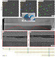
In Figure 3, plan-view and cross-sectional view HRTEM images of the same 1−3-layer MoS2 flake are shown. The high magnification cross-sectional view images in Figures 3f and 3g clearly show that the new layer begins at the bottom for the transition from monolayer to bilayer, as well as from bilayer to tri-layer. This information allows for unambiguous assignment of the substructures in the bi- and tri-layer regions. The S substructure in the monolayer region corresponds to the substructure containing S atoms in the top layer of the bilayer region, as illustrated in Figure 3h. Similarly, the substructure containing S atoms in the top layer of the bilayer region corresponds to the substructure containing S atoms in the outer layers of the tri-layer region. This information allows the defects, identified through the intensity histogram method, to be assigned to the respective substructure, revealing their vertical position.
An atomic column intensity histogram of tri-layer MoS2 is shown in Figure 4a. The (blue) histogram of the substructure containing S atoms in the middle layer shows some defects, but far fewer than the (green) substructure with S atoms in the outer layers. This indicates that although the outer layers provide some protection to the atoms in the middle layer, defects are still created in the middle layer. Single atomic defects in tri-layer MoS2 exhibit a much smaller intensity difference, making them difficult to detect by the naked eye in HRTEM images. Therefore, in Figures 4b–e, alongside HRTEM images of the defects, line scans are presented to compare with HRTEM image simulations. The experimental line fits show good agreement with the simulations, demonstrating the effectiveness of the defect recognition algorithm presented here for up to three layers.
Vertical Position Dependent Damage Cross-Section
Exemplary atomic column intensity histograms of both substructures in 1−3-layer MoS2 are shown in the first two lines of Figure 5. These histograms are presented at the beginning of the image series (filled plot) and for an exemplary higher accumulated dose (bold line). From the atomic column intensity histograms in Figure 5, the separation between the peak corresponding to intact atomic columns and the peak containing defective atomic columns can be determined. The comparison of histograms from different numbers of layers shows that this peak separation decreases as the number of layers increases. This result is expected, as the influence of a missing atom on intensity is reduced when the atomic column contains more atoms. The discussed peak separation, evaluated from defect statistics in single layers of the sample, highlights that, without exploiting the defect statistics, only monolayer MoS2 can be reliably identified, as discussed in [32].
In the intensity histograms shown in Figure 5, the area under the lower intensity peak corresponds to the number of defects, while the area under the higher intensity peak corresponds to the number of intact atomic columns. By comparing the histograms at different accumulated doses (filled plot and bold line), it becomes apparent that, as the electron dose increases, the number of defects increases for all sulfur-containing substructures in 1−3-layer MoS2. Correspondingly, the number of intact atomic columns decreases.
The defect densities as a function of the accumulated dose are plotted in the bottom line of Figure 5. From the slope of this plot, the damage cross-section can be calculated. Therefore, the defect recognition algorithm can efficiently be used to automatically analyze HRTEM image series and determine an experimental value for the damage cross-section. For the monolayer, which contains defects only in one substructure, single and double S vacancies are plotted separately. The obtained damage cross-section of 5.5 barn is consistent with earlier results [12]. Only about 1% of the detected defects are double vacancies, indicating that double vacancies do not significantly impact the evaluation of the damage cross-section. Therefore, for bi- and tri-layer MoS2, single and double vacancies will not be distinguished separately.
Tri-layer MoS2 provides an ideal platform to study defect mechanisms in the middle layer, which is of particular interest because it is shielded from beam damage by the outer layers. In a tri-layer configuration, it is possible to distinguish between defects in the middle layer and those in the outer layers. However, it is not possible to differentiate between defects in the top and bottom layers. The difference in defect creation between the top and bottom layers has already been studied in the bilayer case. Figure 5c shows that upon irradiation with electron beams, defects continuously form in the middle layer of the tri-layer MoS2, which differs from recent work where MoS2 was sandwiched between graphene layers [33].
Resource: Quincke, M., Mundszinger, M., Biskupek, J., & Kaiser, U. (2024). Defect Density and Atomic Defect Recognition in the Middle Layer of a Trilayer MoS2 Stack. Nano Letters, 24(34), 10496–10503. DOI: 10.1021/acs.nanolett.4c02391
-
Chhowalla, M., Shin, H. S., Eda, G., Li, L.-J., Loh, K. P., & Zhang, H. (2013). The chemistry of two-dimensional layered transition metal dichalcogenide nanosheets. Nature Chemistry, 5(4), 263–275. https://doi.org/10.1038/nchem.1589
-
Börner, P. C., Kinyanjui, M. K., Björkman, T., Lehnert, T., Krasheninnikov, A. V., & Kaiser, U. (2018). Observation of charge density waves in free-standing 1T-TaSe2 monolayers by transmission electron microscopy. Applied Physics Letters, 113(17), 173101. https://doi.org/10.1063/1.5052722
-
Stanford, M. G., Pudasaini, P. R., Belianinov, A., Cross, N., Noh, J. H., Koehler, M. R., Mandrus, D. G., Duscher, G., Rondinone, A. J., Ivanov, I. N., Ward, T. Z., & Rack, P. D. (2016). Focused helium-ion beam irradiation effects on electrical transport properties of few-layer WSe2: enabling nanoscale direct write homo-junctions. Scientific Reports, 6(1), 27276. https://doi.org/10.1038/srep27276
-
Keren, I., Dvir, T., Zalic, A., Iluz, A., LeBoeuf, D., Watanabe, K., Taniguchi, T., & Steinberg, H. (2020). Quantum-dot assisted spectroscopy of degeneracy-lifted Landau levels in graphene. Nature Communications, 11(1), 3408. https://doi.org/10.1038/s41467-020-17225-1
-
Dvir, T., Aprili, M., Quay, C. H., & Steinberg, H. (2019). Zeeman tunability of Andreev bound states in van der Waals tunnel barriers. Physical Review Letters, 123(21), 217003. https://doi.org/10.1103/PhysRevLett.123.217003
-
Karnatak, P., Mingazheva, Z., Watanabe, K., Taniguchi, T., Berger, H., Forró, L., & Schönenberger, C. (2023). Origin of subgap states in normal-insulator-superconductor van der Waals heterostructures. Nano Letters, 23(7), 2454–2459. https://doi.org/10.1021/acs.nanolett.2c02777
-
Devidas, T. R., Keren, I., & Steinberg, H. (2021). Spectroscopy of NbSe2 using energy-tunable defect-embedded quantum dots. Nano Letters, 21(16), 6931–6937. https://doi.org/10.1021/acs.nanolett.1c02177
-
Quincke, M., Lehnert, T., Keren, I., Moses Badlyan, N., Port, F., Goncalves, M., Mohn, M. J., Maultzsch, J., Steinberg, H., & Kaiser, U. (2022). Transmission-electron-microscopy-generated atomic defects in two-dimensional nanosheets and their integration in devices for electronic and optical sensing. ACS Applied Nano Materials, 5(8), 11429–11436. https://doi.org/10.1021/acsanm.2c02491
-
Qin, C., Gao, Y., Qiao, Z., Xiao, L., & Jia, S. (2016). Atomic‐layered MoS2 as a tunable optical platform. Advanced Optical Materials, 4(10), 1429–1456. https://doi.org/10.1002/adom.201600323
-
Walker, R. C., Shi, T., Silva, E. C., Jovanovic, I., & Robinson, J. A. (2016). Radiation effects on two‐dimensional materials. Physica Status Solidi (a), 213(12), 3065–3077. https://doi.org/10.1002/pssa.201600395
-
Jiang, J., Xu, T., Lu, J., Sun, L., & Ni, Z. (2019). Defect engineering in 2D materials: Precise manipulation and improved functionalities. Research, 2019, 4641739. https://doi.org/10.34133/2019/4641739
-
Kretschmer, S., Lehnert, T., Kaiser, U., & Krasheninnikov, A. V. (2020). Formation of defects in two-dimensional MoS2 in the transmission electron microscope at electron energies below the knock-on threshold: the role of electronic excitations. Nano Letters, 20(4), 2865–2870. https://doi.org/10.1021/acs.nanolett.0c00670
-
Lehnert, T., Ghorbani-Asl, M., Köster, J., Lee, Z., Krasheninnikov, A. V., & Kaiser, U. (2019). Electron-beam-driven structure evolution of single-layer MoTe2 for quantum devices. ACS Applied Nano Materials, 2(5), 3262–3270. https://doi.org/10.1021/acsanm.9b00616
-
Schleberger, M., & Kotakoski, J. (2018). 2D material science: Defect engineering by particle irradiation. Materials, 11(10), 1885. https://doi.org/10.3390/ma11101885
-
Zhao, X., Kotakoski, J., Meyer, J. C., Sutter, E., Sutter, P., Krasheninnikov, A. V., Kaiser, U., & Zhou, W. (2017). Engineering and modifying two-dimensional materials by electron beams. MRS Bulletin, 42(9), 667–676. https://doi.org/10.1557/mrs.2017.184
-
Liang, Q., Zhang, Q., Zhao, X., Liu, M., & Wee, A. T. (2021). Defect engineering of two-dimensional transition-metal dichalcogenides: applications, challenges, and opportunities. ACS Nano, 15(2), 2165–2181. https://doi.org/10.1021/acsnano.0c09666
-
Srivastava, S., & Mohapatra, Y. N. (2022). Defect density of states in natural and synthetic MoS2 multilayer flakes. Journal of Physics D: Applied Physics, 55(34), 345101. https://doi.org/10.1088/10.1088/1361-6463/ac6f98
-
McDonnell, S., Addou, R., Buie, C., Wallace, R. M., & Hinkle, C. L. (2014). Defect-dominated doping and contact resistance in MoS2. ACS Nano, 8(3), 2880–2888. https://doi.org/10.1021/nn500044q
-
Chakraborty, C., Kinnischtzke, L., Goodfellow, K. M., Beams, R., & Vamivakas, A. N. (2015). Voltage-controlled quantum light from an atomically thin semiconductor. Nature Nanotechnology, 10(6), 507–511. https://doi.org/10.1038/nnano.2015.79
-
Haider, M., Uhlemann, S., Schwan, E., Rose, H., Kabius, B., & Urban, K. (1998). Electron microscopy image enhanced. Nature, 392(6678), 768–769. https://doi.org/10.1038/33823
-
Batson, P. E., Dellby, N., & Krivanek, O. L. (2002). Sub-ångstrom resolution using aberration-corrected electron optics. Nature, 418(6898), 617–620. https://doi.org/10.1038/nature00972
-
Köster, J., Storm, A., Ghorbani-Asl, M., Kretschmer, S., Gorelik, T. E., Krasheninnikov, A. V., & Kaiser, U. (2022). Structural and chemical modifications of few-layer transition metal phosphorous trisulfides by electron irradiation. The Journal of Physical Chemistry C, 126(36), 15446–15455. https://doi.org/10.1021/acs.jpcc.2c03800
-
Linck, M., Hartel, P., Uhlemann, S., Kahl, F., Müller, H., Zach, J., Haider, M., Niestadt, M., Bischoff, M., Biskupek, J., Lee, Z., Lehnert, T., Börrnert, F., Rose, H., & Kaiser, U. (2016). Chromatic aberration correction for atomic resolution TEM imaging from 20 to 80 kV. Physical Review Letters, 117(7), 076101. https://doi.org/10.1103/PhysRevLett.117.076101
-
Komsa, H. P., Kotakoski, J., Kurasch, S., Lehtinen, O., Kaiser, U., & Krasheninnikov, A. V. (2012). Two-dimensional transition metal dichalcogenides under electron irradiation: Defect production and doping. Physical Review Letters, 109(3), 035503. https://doi.org/10.1103/PhysRevLett.109.035503
-
Leist, C., He, M., Liu, X., Kaiser, U., & Qi, H. (2022). Deep-learning pipeline for statistical quantification of amorphous two-dimensional materials. ACS Nano, 16(12), 20488–20496. https://doi.org/10.1021/acsnano.2c06807
-
Yang, S. H., Choi, W., Cho, B. W., Agyapong-Fordjour, F. O. T., Park, S., Yun, S. J., Kim, H.-J., Han, Y.-K., Lee, Y. H., Kim, K. K., & Kim, Y. M. (2021). Deep learning-assisted quantification of atomic dopants and defects in 2D materials. Advanced Science, 8(16), 2101099. https://doi.org/10.1002/advs.202101099
-
Nord, M., Vullum, P. E., MacLaren, I., Tybell, T., & Holmestad, R. (2017). Atomap: a new software tool for the automated analysis of atomic resolution images using two-dimensional Gaussian fitting. Advanced Structural and Chemical Imaging, 3, 1–12. https://doi.org/10.1186/s40679-017-0042-5
-
De Backer, A., Van den Bos, K. H. W., Van den Broek, W., Sijbers, J., & Van Aert, S. (2016). StatSTEM: An efficient approach for accurate and precise model-based quantification of atomic resolution electron microscopy images. Ultramicroscopy, 171, 104–116. https://doi.org/10.1016/j.ultramic.2016.08.018
-
Mukherjee, D., Miao, L., Stone, G., & Alem, N. (2020). Mpfit: A robust method for fitting atomic resolution images with multiple Gaussian peaks. Advanced Structural and Chemical Imaging, 6, 1–12. https://doi.org/10.1186/s40679-020-0068-y
-
Momma, K., & Izumi, F. (2011). VESTA 3 for three-dimensional visualization of crystal, volumetric and morphology data. Journal of Applied Crystallography, 44(6), 1272–1276. https://doi.org/10.1107/S0021889811038970
-
Madsen, J., & Susi, T. (2021). The abTEM code: transmission electron microscopy from first principles. Open Research Europe, 1, 24. https://doi.org/10.12688/openreseurope.13015.2
-
Köster, J., Storm, A., Gorelik, T. E., Mohn, M. J., Port, F., Gonçalves, M. R., & Kaiser, U. (2022). Evaluation of TEM methods for their signature of the number of layers in mono- and few-layer TMDs as exemplified by MoS2 and MoTe2. Micron, 160, 103303. https://doi.org/10.1016/j.micron.2022.103303
-
Lehnert, T., Lehtinen, O., Algara-Siller, G., & Kaiser, U. (2017). Electron radiation damage mechanisms in 2D MoSe2. Applied Physics Letters, 110(3), 033102. https://doi.org/10.1063/1.4973809


